Epithelial Cell Adaptation on Nanoporous Titanium Surfaces
Yannis Karamitsos, Dainelys Guadarrama Bello, Audrey Brodeur, Antonio Nanci
Laboratory for the Study of Calcified Tissues and Biomaterials, Faculté de Médecine Dentaire, Université de Montréal
The existing literature presents divergent results regarding the optimal surface texturization state for ensuring peri-implant tissue health. While the influence of nanoscale surface modifications on
osteogenic cells has been well acknowledged, the impact on epithelial cell behavior warrants further exploration. This study investigates epithelial cell response to two distinct titanium surfaces
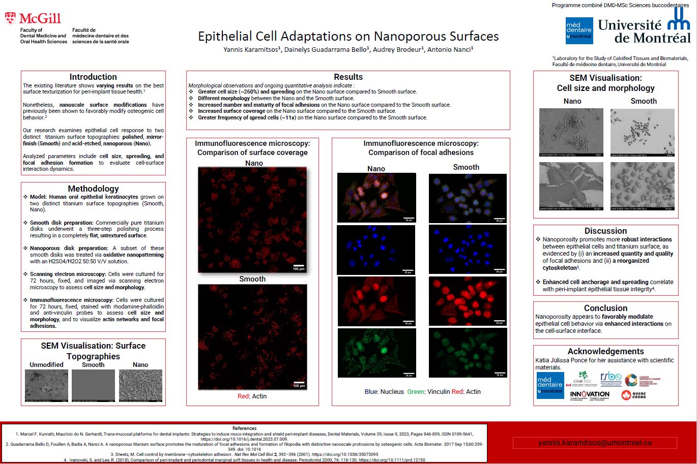
topographies: a completely smooth, “mirror-finish” control surface and an acid-etched, nanoporous surface which has previously demonstrated efficient modulation of bone cell response. We assess cell size, spreading, and formation of focal adhesions as a means of evaluating cellsurface interaction dynamics.
In our methodology, human oral epithelial cells were utilised as a model to study peri-implant epithelial interaction with titanium surfaces. Commercially pure titanium disks underwent a threestep polishing process, resulting in a completely smooth surface, establishing a control baseline
free of any macro- or micro- scale texturization. A subset of these smooth disks underwent oxidative nanopatterning with a solution of H2SO4/H2O2 50:50 V/V. Cells were cultured on these surfaces for 72 hours, followed by fixation in periodate L-lysine paraformaldehyde and staining with rhodamine-phalloidin and anti-vinculin probes to visualize actin networks and focal adhesions. Additional samples were fixed with osmium, dehydrated with alcohol, and examined using scanning electron microscopy.
Our findings, from both scanning electron microscopy and fluorescence microscopy, revealed that cells on the nanoporous surface not only exhibit markedly greater spreading and size compared to those on the polished surface, suggesting more robust cell-surface interactions, but also display a significantly different morphology—transitioning from round on smooth surfaces to spread-out and polygonal on the nanoporous surface. A notable increase in the number and maturity of focal adhesions on the nanoporous surface further underscores its role in enhancing cell anchorage.
In summary, nanoporosity appears to favorably modulate epithelial cell behavior, suggesting enhanced interactions on the cell-surface interface. Future investigations should extend to the influence of nanoporosity on other aspects of epithelial cell function, including replication and adhesion strength, and assess the overall integrity and health of the peri-implant epithelium through in vivo studies.