Materials Research is an interdisciplinary subject bringing many traditional sciences together with engineering, in the investigation of the relationships between and among the structure and properties of materials, and how these related to materials processing and performance. MMC is a multi-user, shared instrumentation facilities for materials research and characterization. It regroups various laboratories:
MMC-Analytical Laboratories
Agilent MP-AES Microwave Plasma Atomic Emission Spectroscopy
- Simultaneous multi-analyte determination of major and minor elements. (ppm/ppb level)
- Samples must be in solution and free from particulates.
Micromeritics TriStar 3000 Surface area and porosity analyzer
- BET analysis of up to 3 samples at the time
- Porosity
Bio Tek Synergy H1 Hybrid Multi-Mode Microplate Reader
- Hybrid multi-mode microplate reader with the following detection modes:
- Fluorescence (250-700 nm),
- Luminescence (300-700 nm)
- UV-visible absorbance (230-999 nm)
Microtrac Flow Sync wet laser diffraction
- size distribution of particles and particle’s shape data in a liquid medium
- Size range from 0.020 to 2000 microns
Malvern Zetasizer Nano ZS
- Ability to measure three fundamental characteristics of particles or molecules in a liquid medium
- Particle size (0.3nm-10 microns)
- Zeta potential
- Molecular weight
Easypure RF Compact ultrapure and MicroPure water system
- Produces ultrapure water (deionized water)
Perkin Elmer Lamda 20 UV/Vis Spectrometer
Dionex DX-00 Ion Chromatography
MMC-Materials Laboratories
 |
|
MMC-Characterization Laboratory
Stereo zoom microscope-Olympus SZ40
- 067x-4x
- Sony color video camera
- Clemex Vision image acquisition and analysis software
Inverted metallurgical optical microscope-Nikon Epiphot 200
- 25x-1000x
- Automatic stage
- Clemex Vision image acquisition and analysis software
Digital microscope Keyence VHX500
- Characterization tool exceeding the limits of optical observation (high camera frame rate, fully focused image)
- 20x-2000x, 3D observation, 2D and 3D measurement tools and stitching capabilities (Mosaic), large depth of field and 360° free-angle observation
- Various lighting
- Quantitative metallography and Image analysis
Bruker D8 Discovery X-Ray Diffractometer (Cobalt source
 |
|
MMC-Electron Microscopy Laboratories
Hitachi Tabletop microscope TM4000
- Backscattered electron
- Magnifications : x10-x100,000(Photographic magnification) x25-x250,000(Monitor display magnification)
- Vacuum mode: BSE standard and charge-up reduction
- Accelerating voltage: 5kV,10kV and 15kV
Hitachi SU-3500 Variable Pressure -SEM
 |
|
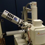
Hitachi Cold FE SU-8000 SEM
 |
|
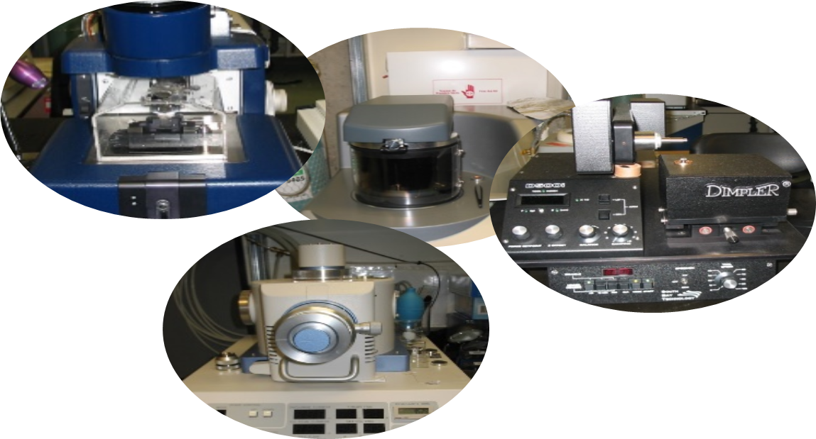
Other equipment
The Department of Mining and Materials Engineering, McGill University has experts in a variety of materials fields ranging from aerospace, automotive, biomaterials and tissue engineering, characterization, electron microscopy, computer-aided modelling, electronic materials, energy, hydrothermal processing, metal processing, mineral processing, nano materials, pyrometallurgy, and surface science and engineering. We characterize materials, we study with state-of-the-art technology, and we model their properties with the help of computers.
The list of equipment includes some instruments that are not operated by MMC but by individual researchers or other shared research facilities. You can contact us if you are looking for a specific instrument. MMC will direct you to the appropriate person for your characterization work.
McGill Electron Microscopy Research Group (MEMRG)
Professor Gauvin. Mining and Materials Engineering Department
https://www.memrg.com/our-equipment
Facility for Electron Microscopy Research (FEMR)
https://www.mcgill.ca/femr/instrumentation